How is Acute Myeloid Leukemia Diagnosed?
Acute myeloid leukemia (AML) is diagnosed through a series of tests that examine the blood and bone marrow. The process usually begins with a physical examination where the doctor checks for physical signs of AML such as pale skin, fatigue, bruising and shortness of breath. If AML is suspected, the following tests may be conducted:
Blood Tests:
- Complete blood count (CBC): The complete blood count (CBC) is a test that measures the levels of red blood cells, white blood cells, and platelets in the blood. If leukemia cells occupy too much of the bone marrow, levels of red blood cells, white blood cells and platelets will be low and anemia may be present.
- Complete metabolic panel (CMP): The complete metabolic panel (CMP) is a test that measures the levels of electrolytes in the blood. It also looks at markers like BUN and creatinine to assess kidney function and markers like AST and ALT to assess liver function. High levels of any of these labs can indicate that the kidneys or liver are not functioning as well as they should.
Bone Marrow Tests:
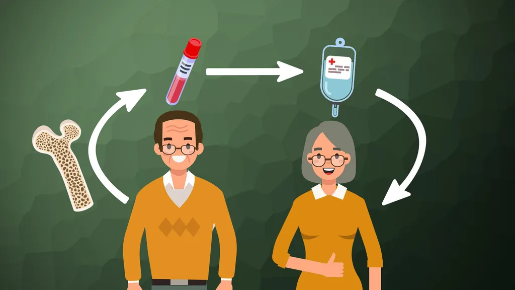
- Bone marrow biopsy and aspiration: This is the most invasive of the diagnostic tests but the information gathered is extremely valuable. In this procedure, the back of the pelvic bone is numbed with a local anesthetic. For the aspiration, a needle is inserted into the bone, and a syringe is used to remove a small amount of liquid bone marrow. This causes a brief, sharp pain. Then for the biopsy, a needle is used to remove a tiny sliver of bone and marrow, about 1/16-inch across and 1-inch long. There may be some soreness in the biopsy area when the numbing medicine wears off. Most patients go home immediately after the procedure. If you prefer, the procedure can be done under light anesthesia called conscious sedation. The sedation makes you tired for an hour or two but doesn’t knock you out. Many patients prefer to go without any pain medication. There is no “wrong” decision.
- Immunohistochemistry: In this test, a part of the bone marrow biopsy sample is treated with special antibodies (man-made versions of immune system proteins) that attach only to specific molecules on the cell surface. These antibodies cause color changes, which can be seen under a microscope. This test may be helpful in differentiating between cell types and in finding leukemia cells.
- Flow cytometry: Like immunohistochemistry, this test looks for certain substances on the outside surface of cells that help identify what types of cells they are. This test is able to look at many more cells than immunohistochemistry. For this test, a sample of cells is treated with special antibodies that stick to the cells only if certain substances are present on their surfaces. The cells are then passed in front of a laser beam. If the cells now have antibodies attached to them, the laser will cause them to give off light, which can be measured and analyzed by a computer. Groups of cells can be separated and counted by these methods. This is the most commonly used test for immunophenotyping, the process of classifying cells according to the substances (antigens) on their surfaces. Different cells and cell types have different antigens on their surface. These antigens may also change as each cell matures. Flow cytometry can help determine if there are abnormal cells in the bone marrow and if they are leukemia cells, AML cells, lymphoma cells, some other cancer, or a non-cancerous disease.
- Karyotyping: This technique allows doctors to evaluate the chromosomes (long strands of DNA) in normal bone marrow cells and leukemia cells. The cells are examined under a microscope to see if the chromosomes have any abnormalities such as a translocation. Translocations occur when part of one chromosome has broken off and becomes attached to another chromosome. This can happen in some cases of AML. Some leukemia cells may have too many chromosomes, too few chromosomes, or other chromosome abnormalities. Finding these changes can sometimes help predict prognosis and determine treatment options.
- Fluorescent in situ hybridization (FISH): Fluorescent in situ hybridization (FISH) is similar to cytogenetic testing. It uses special fluorescent dyes that only attach to specific parts of chromosomes. FISH can find most chromosome changes (such as translocations) that can be seen under a microscope in standard cytogenetic tests, as well as some changes too small to be seen with usual cytogenetic testing. FISH can be used to look for specific changes in chromosomes. It can be used on regular blood or bone marrow samples. It is very accurate and can usually provide results within a couple of days, which is why this test is now used in many medical centers.
- Next generation sequencing (NGS): Using a bone marrow biopsy sample, the leukemia cells are purified and the genetic material is extracted. This test provides everything that the FISH test provides in terms of translocations, but also identifies gene “signatures” or genes that are turned on or off, or are over or under-expressed. This provides redundant information from the FISH but looks at leukemia on a molecular level and can test for 35,000 genes in a single test. This test provides valuable genetic information about the subtype of AML, allowing patients to receive more personalized treatments.
Imaging Tests
- Computed tomography (CT) scan: The computed tomography (CT) scan (also known as a CAT scan) is an X-ray procedure that produces detailed cross-sectional images of your body. Instead of taking one picture, like a conventional X-ray, a CT scanner takes many pictures of the part of the body being studied as it rotates. A CT scan can show enlarged lymph nodes, a swollen spleen, or pockets of infection in your organs. Oftentimes, a patient will drink 1 to 2 pints of a solution of contrast material before the scan. This helps outline the intestine so that it is not mistaken for tumors. A patient may also receive an intravenous (IV) line through which a different contrast dye is injected. This helps better outline body structures. The injection can cause a feeling of warmth throughout the body (flushing). Some people are allergic to the IV contrast and should tell their doctor if they have ever had a reaction to any contrast material used for X-rays. CT scans take longer than regular X-rays and require lying motionless on a table while images are taken. Some patients might feel a bit confined during the scan, but the tests are over relatively quickly. CT scans can also be used to guide a biopsy needle precisely into a suspected tumor. For this procedure, called a CT-guided needle biopsy, the patient remains on the CT scanning table while a radiologist advances a biopsy needle toward the location of the tumor. CT scans are repeated until the needle is within the mass. A fine needle biopsy sample or a core needle biopsy sample is removed and examined under a microscope.
- Magnetic resonance imaging (MRI) scan: MRI scans use radio waves and strong magnets instead of X-rays. The energy from the radio waves is absorbed and then released in a pattern formed by the type of tissue and by certain diseases. A computer translates the pattern of radio waves given off by the tissues into a very detailed image of parts of the body. Not only does this produce cross-sectional slices of the body like a CT scanner, but it can also produce slices that are parallel with the length of your body. A dye might be injected just as with CT scans but is used less often. This test may be used to see if leukemia has spread to the brain or spinal cord. MRI scans are a little more uncomfortable than CT scans. They can take an hour or longer, and the patient is placed inside tunnel-like equipment, which can feel confining. The machine makes a thumping noise but the facility can provide headphones with music to block it out.
- Positron emission tomography (PET) scan: With a PET scan, radioactive glucose (sugar) is injected into the patient’s vein to look for cancer cells. Because cancers use glucose (sugar) at a higher rate than normal tissues, the radioactivity will tend to concentrate in the cancer. A scanner is used to spot radioactive deposits.
A Summary of Diagnosing Acute Myeloid Leukemia
It's important to note that the diagnosis and classification of AML is complex and requires a comprehensive evaluation by a team of specialists. A complete diagnosis requires a physical examination, looking at a bone marrow sample under the microscope, immunophenotyping, karyotype analysis and molecular genetic testing. The specific markers and genetic changes found in the bone marrow sample can also help determine a person's prognosis and guide treatment decisions. If you don't currently have an AML specialist on your team, it is important that you consult with one. Use HealthTree's AML Specialist Directory to locate a specialist near you.
Want to Learn More About Acute Myeloid Leukemia?
Keep reading HealthTree for Acute Myeloid Leukemia's 101 pages!
Acute myeloid leukemia (AML) is diagnosed through a series of tests that examine the blood and bone marrow. The process usually begins with a physical examination where the doctor checks for physical signs of AML such as pale skin, fatigue, bruising and shortness of breath. If AML is suspected, the following tests may be conducted:
Blood Tests:
- Complete blood count (CBC): The complete blood count (CBC) is a test that measures the levels of red blood cells, white blood cells, and platelets in the blood. If leukemia cells occupy too much of the bone marrow, levels of red blood cells, white blood cells and platelets will be low and anemia may be present.
- Complete metabolic panel (CMP): The complete metabolic panel (CMP) is a test that measures the levels of electrolytes in the blood. It also looks at markers like BUN and creatinine to assess kidney function and markers like AST and ALT to assess liver function. High levels of any of these labs can indicate that the kidneys or liver are not functioning as well as they should.
Bone Marrow Tests:
- Bone marrow biopsy and aspiration: This is the most invasive of the diagnostic tests but the information gathered is extremely valuable. In this procedure, the back of the pelvic bone is numbed with a local anesthetic. For the aspiration, a needle is inserted into the bone, and a syringe is used to remove a small amount of liquid bone marrow. This causes a brief, sharp pain. Then for the biopsy, a needle is used to remove a tiny sliver of bone and marrow, about 1/16-inch across and 1-inch long. There may be some soreness in the biopsy area when the numbing medicine wears off. Most patients go home immediately after the procedure. If you prefer, the procedure can be done under light anesthesia called conscious sedation. The sedation makes you tired for an hour or two but doesn’t knock you out. Many patients prefer to go without any pain medication. There is no “wrong” decision.
- Immunohistochemistry: In this test, a part of the bone marrow biopsy sample is treated with special antibodies (man-made versions of immune system proteins) that attach only to specific molecules on the cell surface. These antibodies cause color changes, which can be seen under a microscope. This test may be helpful in differentiating between cell types and in finding leukemia cells.
- Flow cytometry: Like immunohistochemistry, this test looks for certain substances on the outside surface of cells that help identify what types of cells they are. This test is able to look at many more cells than immunohistochemistry. For this test, a sample of cells is treated with special antibodies that stick to the cells only if certain substances are present on their surfaces. The cells are then passed in front of a laser beam. If the cells now have antibodies attached to them, the laser will cause them to give off light, which can be measured and analyzed by a computer. Groups of cells can be separated and counted by these methods. This is the most commonly used test for immunophenotyping, the process of classifying cells according to the substances (antigens) on their surfaces. Different cells and cell types have different antigens on their surface. These antigens may also change as each cell matures. Flow cytometry can help determine if there are abnormal cells in the bone marrow and if they are leukemia cells, AML cells, lymphoma cells, some other cancer, or a non-cancerous disease.
- Karyotyping: This technique allows doctors to evaluate the chromosomes (long strands of DNA) in normal bone marrow cells and leukemia cells. The cells are examined under a microscope to see if the chromosomes have any abnormalities such as a translocation. Translocations occur when part of one chromosome has broken off and becomes attached to another chromosome. This can happen in some cases of AML. Some leukemia cells may have too many chromosomes, too few chromosomes, or other chromosome abnormalities. Finding these changes can sometimes help predict prognosis and determine treatment options.
- Fluorescent in situ hybridization (FISH): Fluorescent in situ hybridization (FISH) is similar to cytogenetic testing. It uses special fluorescent dyes that only attach to specific parts of chromosomes. FISH can find most chromosome changes (such as translocations) that can be seen under a microscope in standard cytogenetic tests, as well as some changes too small to be seen with usual cytogenetic testing. FISH can be used to look for specific changes in chromosomes. It can be used on regular blood or bone marrow samples. It is very accurate and can usually provide results within a couple of days, which is why this test is now used in many medical centers.
- Next generation sequencing (NGS): Using a bone marrow biopsy sample, the leukemia cells are purified and the genetic material is extracted. This test provides everything that the FISH test provides in terms of translocations, but also identifies gene “signatures” or genes that are turned on or off, or are over or under-expressed. This provides redundant information from the FISH but looks at leukemia on a molecular level and can test for 35,000 genes in a single test. This test provides valuable genetic information about the subtype of AML, allowing patients to receive more personalized treatments.
Imaging Tests
- Computed tomography (CT) scan: The computed tomography (CT) scan (also known as a CAT scan) is an X-ray procedure that produces detailed cross-sectional images of your body. Instead of taking one picture, like a conventional X-ray, a CT scanner takes many pictures of the part of the body being studied as it rotates. A CT scan can show enlarged lymph nodes, a swollen spleen, or pockets of infection in your organs. Oftentimes, a patient will drink 1 to 2 pints of a solution of contrast material before the scan. This helps outline the intestine so that it is not mistaken for tumors. A patient may also receive an intravenous (IV) line through which a different contrast dye is injected. This helps better outline body structures. The injection can cause a feeling of warmth throughout the body (flushing). Some people are allergic to the IV contrast and should tell their doctor if they have ever had a reaction to any contrast material used for X-rays. CT scans take longer than regular X-rays and require lying motionless on a table while images are taken. Some patients might feel a bit confined during the scan, but the tests are over relatively quickly. CT scans can also be used to guide a biopsy needle precisely into a suspected tumor. For this procedure, called a CT-guided needle biopsy, the patient remains on the CT scanning table while a radiologist advances a biopsy needle toward the location of the tumor. CT scans are repeated until the needle is within the mass. A fine needle biopsy sample or a core needle biopsy sample is removed and examined under a microscope.
- Magnetic resonance imaging (MRI) scan: MRI scans use radio waves and strong magnets instead of X-rays. The energy from the radio waves is absorbed and then released in a pattern formed by the type of tissue and by certain diseases. A computer translates the pattern of radio waves given off by the tissues into a very detailed image of parts of the body. Not only does this produce cross-sectional slices of the body like a CT scanner, but it can also produce slices that are parallel with the length of your body. A dye might be injected just as with CT scans but is used less often. This test may be used to see if leukemia has spread to the brain or spinal cord. MRI scans are a little more uncomfortable than CT scans. They can take an hour or longer, and the patient is placed inside tunnel-like equipment, which can feel confining. The machine makes a thumping noise but the facility can provide headphones with music to block it out.
- Positron emission tomography (PET) scan: With a PET scan, radioactive glucose (sugar) is injected into the patient’s vein to look for cancer cells. Because cancers use glucose (sugar) at a higher rate than normal tissues, the radioactivity will tend to concentrate in the cancer. A scanner is used to spot radioactive deposits.
A Summary of Diagnosing Acute Myeloid Leukemia
It's important to note that the diagnosis and classification of AML is complex and requires a comprehensive evaluation by a team of specialists. A complete diagnosis requires a physical examination, looking at a bone marrow sample under the microscope, immunophenotyping, karyotype analysis and molecular genetic testing. The specific markers and genetic changes found in the bone marrow sample can also help determine a person's prognosis and guide treatment decisions. If you don't currently have an AML specialist on your team, it is important that you consult with one. Use HealthTree's AML Specialist Directory to locate a specialist near you.
Want to Learn More About Acute Myeloid Leukemia?
Keep reading HealthTree for Acute Myeloid Leukemia's 101 pages!
Trending Articles


Get the latest thought leadership on your Acute Myeloid Leukemia delivered straight to your inbox
Subscribe to the weekly newsletter for news, stories, clinical trial updates, and helpful resources and events with cancer experts.
Follow Us


HealthTree Foundation is a qualified 501(c)(3) tax-exempt organization.
Copyright © 2024 HealthTree Foundation. All rights reserved.
Tax ID 45-5354811