What Makes it CLL? Looking at Chromosome Deletions

A CLL cell is a mutated B-cell that is not capable of doing its normal job of defending the body against invaders like bacteria or viruses. The instructions that tell the cell to die are changed so the ineffective B-cell keeps multiplying.
This happens because certain parts of the B-cell’s instructions (chromosomes) that tell the cell how to work are deleted or mutated.

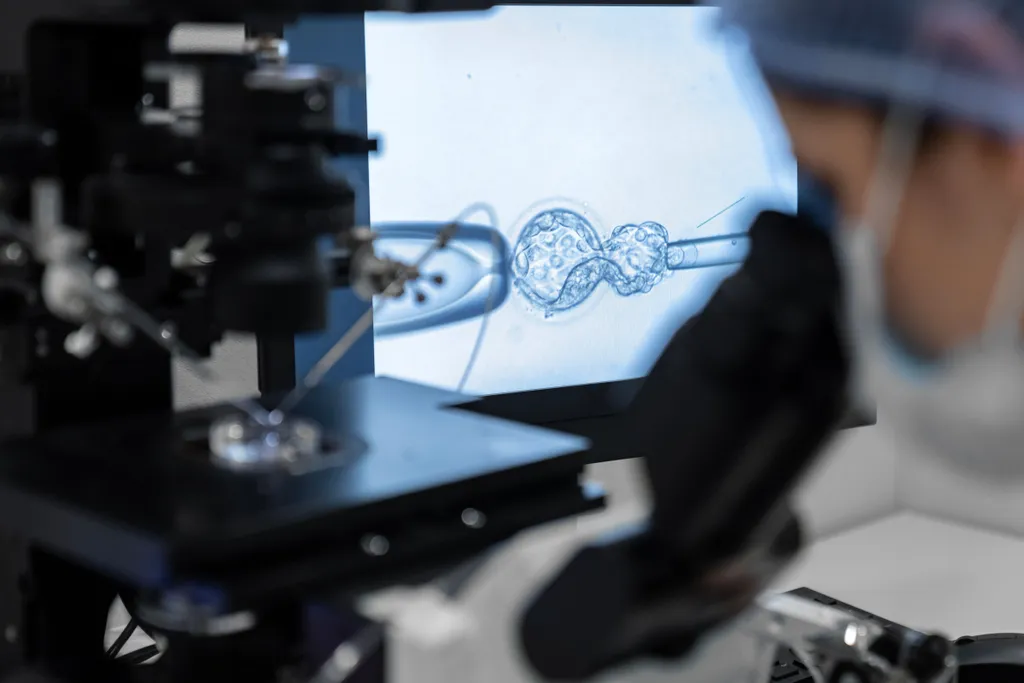
To check for chromosome deletions, a doctor will complete a fluorescence in situ hybridization (FISH) test.
- A FISH test is completed by collecting a blood sample from the CLL patient which includes a set of the patient’s full chromosomes. The sample is placed on a glass slide and exposed to a small piece of purified DNA tagged with a fluorescent dye (probe). The fluorescently labeled probe finds and then binds to its matching sequence within the set of chromosomes. With the use of a special microscope, the chromosome location where the fluorescent probe bound can be seen (genome.gov)

(Image: genome.gov)
The B-cell will be labeled as CLL if part of chromosomes 11q, 13q, or 17p are deleted. Some cells may also have an extra copy of chromosome 12 (q = long arm of the chromosome, p= short arm of the chromosome).

The location and size of the deletion in each chromosome may vary among patients. Patients should create a care plan based on their individual situation with a CLL specialist.
Need Help Finding a CLL Specialist?
Check out HealthTree’s CLL Specialist Directory.
A CLL cell is a mutated B-cell that is not capable of doing its normal job of defending the body against invaders like bacteria or viruses. The instructions that tell the cell to die are changed so the ineffective B-cell keeps multiplying.
This happens because certain parts of the B-cell’s instructions (chromosomes) that tell the cell how to work are deleted or mutated.
To check for chromosome deletions, a doctor will complete a fluorescence in situ hybridization (FISH) test.
- A FISH test is completed by collecting a blood sample from the CLL patient which includes a set of the patient’s full chromosomes. The sample is placed on a glass slide and exposed to a small piece of purified DNA tagged with a fluorescent dye (probe). The fluorescently labeled probe finds and then binds to its matching sequence within the set of chromosomes. With the use of a special microscope, the chromosome location where the fluorescent probe bound can be seen (genome.gov)
(Image: genome.gov)
The B-cell will be labeled as CLL if part of chromosomes 11q, 13q, or 17p are deleted. Some cells may also have an extra copy of chromosome 12 (q = long arm of the chromosome, p= short arm of the chromosome).
The location and size of the deletion in each chromosome may vary among patients. Patients should create a care plan based on their individual situation with a CLL specialist.
Need Help Finding a CLL Specialist?
Check out HealthTree’s CLL Specialist Directory.

about the author
Megan Heaps
Megan joined HealthTree in 2022. She enjoys helping patients and their care partners understand the various aspects of the cancer. This understanding enables them to better advocate for themselves and improve their treatment outcomes.
More on Navigating Your Health


Trending Articles


Get the Latest Chronic Lymphocytic Leukemia Updates, Delivered to You.
By subscribing to the HealthTree newsletter, you'll receive the latest research, treatment updates, and expert insights to help you navigate your health.
Together we care.
Together we cure.