Tests for CLL Patients: IHC on Biopsy Sample

CLL patients should receive an immunohistochemistry (IHC) on biopsy sample test as part of their diagnosis. IHC is a technique used in medical diagnostics and research to study various diseases, including CLL. It involves the use of specific antibodies to detect and visualize specific proteins or molecules within tissue samples obtained through a biopsy. By providing important insights into cellular and molecular composition, IHC plays a crucial role in understanding CLL at a microscopic level.
Understanding Immunohistochemistry (IHC)
Immunohistochemistry (IHC) is a technique that combines the principles of immunology (the study of the immune system) and histology (the study of tissues). It allows scientists and doctors to visualize specific proteins or molecules within tissues by using antibodies that bind to their targets. These targets can be cancer-specific markers, signaling molecules, or other proteins of interest.
What is a Biopsy?
A biopsy is a medical procedure in which a small piece of tissue or cells is removed from a patient for further review. For CLL patients, this may include reviewing a lymph node, a section of bone marrow, or a small blood sample.
How does IHC work?
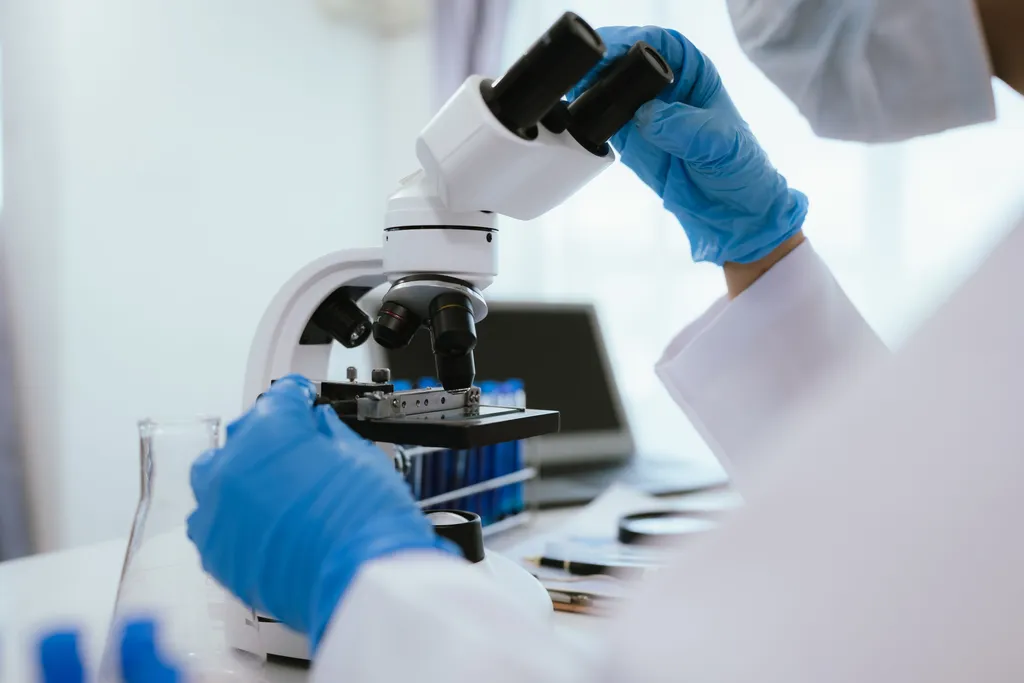
- Sample Preparation: The first step in IHC is to obtain a tissue sample through a biopsy procedure (small blood sample, bone marrow sample, and/or lymph node). The sample is then treated and processed to create thin sections for microscopic analysis.
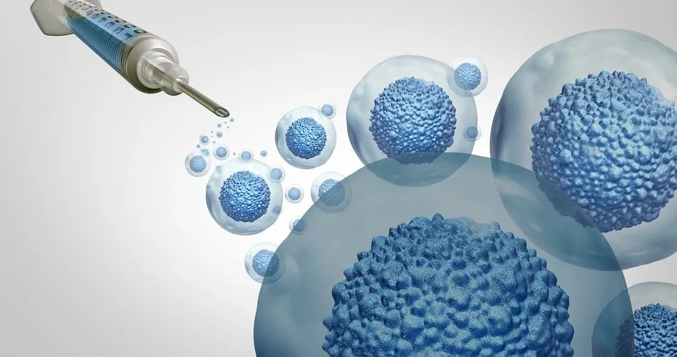
- Primary Antibodies: Next, primary antibodies are selected. Primary antibodies are specific proteins that recognize and bind to the target molecules of interest. These antibodies can be commercially available or specifically developed for a particular study.
- Antibody Incubation: The tissue sections are incubated with the primary antibodies. If the target molecule is present in the tissue, the primary antibodies will bind to it, forming an antibody-antigen complex.
- Detection Systems: To visualize the antibody-antigen complex, a detection system is used. This system can be based on enzymes, fluorescent dyes, or nanoparticles. These systems generate a visible signal that can be detected under a microscope.
- Visualization: The tissue sections are examined under a microscope. If the target molecule is present, the antibody-antigen complex will be visible as a colored stain or fluorescent signal. The intensity and location of the stain provide information about the presence, abundance, and cellular localization of the target molecule.
Applications of IHC
- Cancer Diagnosis: IHC helps identify specific markers expressed in CLL cells to help accurately diagnose CLL. The genetic markers IHC looks for in CLL include surface proteins CD5, CD19, CD20, and CD23 (see here for more about CLL surface proteins)
- Research and Medicine Development: IHC enables researchers to study the expression patterns and distribution of specific proteins in tissues, contributing to a better understanding of disease mechanisms and the development of targeted therapies
CLL patients should receive an immunohistochemistry (IHC) on biopsy sample test as part of their diagnosis. IHC is a technique used in medical diagnostics and research to study various diseases, including CLL. It involves the use of specific antibodies to detect and visualize specific proteins or molecules within tissue samples obtained through a biopsy. By providing important insights into cellular and molecular composition, IHC plays a crucial role in understanding CLL at a microscopic level.
Understanding Immunohistochemistry (IHC)
Immunohistochemistry (IHC) is a technique that combines the principles of immunology (the study of the immune system) and histology (the study of tissues). It allows scientists and doctors to visualize specific proteins or molecules within tissues by using antibodies that bind to their targets. These targets can be cancer-specific markers, signaling molecules, or other proteins of interest.
What is a Biopsy?
A biopsy is a medical procedure in which a small piece of tissue or cells is removed from a patient for further review. For CLL patients, this may include reviewing a lymph node, a section of bone marrow, or a small blood sample.
How does IHC work?
- Sample Preparation: The first step in IHC is to obtain a tissue sample through a biopsy procedure (small blood sample, bone marrow sample, and/or lymph node). The sample is then treated and processed to create thin sections for microscopic analysis.
- Primary Antibodies: Next, primary antibodies are selected. Primary antibodies are specific proteins that recognize and bind to the target molecules of interest. These antibodies can be commercially available or specifically developed for a particular study.
- Antibody Incubation: The tissue sections are incubated with the primary antibodies. If the target molecule is present in the tissue, the primary antibodies will bind to it, forming an antibody-antigen complex.
- Detection Systems: To visualize the antibody-antigen complex, a detection system is used. This system can be based on enzymes, fluorescent dyes, or nanoparticles. These systems generate a visible signal that can be detected under a microscope.
- Visualization: The tissue sections are examined under a microscope. If the target molecule is present, the antibody-antigen complex will be visible as a colored stain or fluorescent signal. The intensity and location of the stain provide information about the presence, abundance, and cellular localization of the target molecule.
Applications of IHC
- Cancer Diagnosis: IHC helps identify specific markers expressed in CLL cells to help accurately diagnose CLL. The genetic markers IHC looks for in CLL include surface proteins CD5, CD19, CD20, and CD23 (see here for more about CLL surface proteins)
- Research and Medicine Development: IHC enables researchers to study the expression patterns and distribution of specific proteins in tissues, contributing to a better understanding of disease mechanisms and the development of targeted therapies

about the author
Megan Heaps
Megan joined HealthTree in 2022. She enjoys helping patients and their care partners understand the various aspects of the cancer. This understanding enables them to better advocate for themselves and improve their treatment outcomes.
More on Navigating Your Health


Trending Articles


Get the Latest Chronic Lymphocytic Leukemia Updates, Delivered to You.
By subscribing to the HealthTree newsletter, you'll receive the latest research, treatment updates, and expert insights to help you navigate your health.
Together we care.
Together we cure.