What Makes it CLL? A Look at Membrane Proteins

To check for CLL, one of the things a patient’s doctor will look for are certain proteins on the surface of the B-cell’s membrane. Different cancers will have a common protein pattern on the cell’s surface (immunophenotype). The B-cell will be labeled as CLL if proteins CD5, CD19, and CD23 are on the B-cell’s surface. Some CLL cells may also have surface protein CD20. To classify as CLL, the cells should never have surface protein CD10.
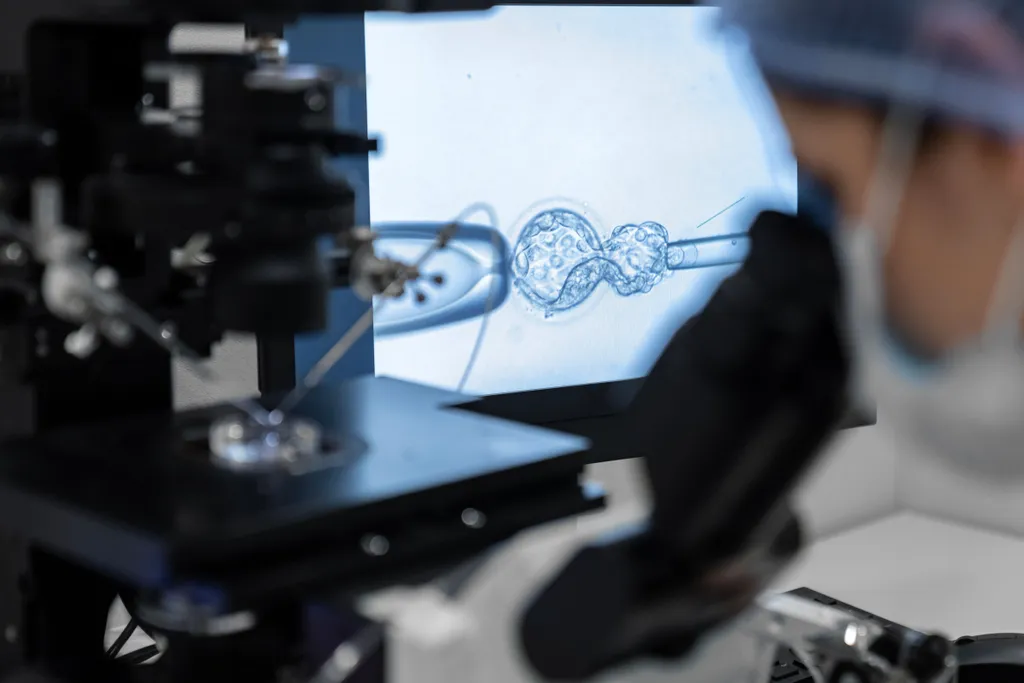
To check for the protein pattern, the CLL patient’s doctor will complete a flow cytometry test. This is done by taking a sample of the patient’s blood and mixing it with a solution that includes fluorescent dyed antibodies to attach to each of the CLL cell’s specific proteins (CD5, CD19, CD20, and CD23). The sample is placed into the flow cytometer machine and the cells move single file past a laser. As the laser shines on each cell, the fluorescent antibody markers that have attached to the cell’s proteins will illuminate their dyed color. The computer records information from the test.

(Image source: Abbexa)
A diagnosis of CLL is made if the test results show there are at least 5,000 copies of the same CLL cell.
To check for CLL, one of the things a patient’s doctor will look for are certain proteins on the surface of the B-cell’s membrane. Different cancers will have a common protein pattern on the cell’s surface (immunophenotype). The B-cell will be labeled as CLL if proteins CD5, CD19, and CD23 are on the B-cell’s surface. Some CLL cells may also have surface protein CD20. To classify as CLL, the cells should never have surface protein CD10.
To check for the protein pattern, the CLL patient’s doctor will complete a flow cytometry test. This is done by taking a sample of the patient’s blood and mixing it with a solution that includes fluorescent dyed antibodies to attach to each of the CLL cell’s specific proteins (CD5, CD19, CD20, and CD23). The sample is placed into the flow cytometer machine and the cells move single file past a laser. As the laser shines on each cell, the fluorescent antibody markers that have attached to the cell’s proteins will illuminate their dyed color. The computer records information from the test.
(Image source: Abbexa)
A diagnosis of CLL is made if the test results show there are at least 5,000 copies of the same CLL cell.

about the author
Megan Heaps
Megan joined HealthTree in 2022. She enjoys helping patients and their care partners understand the various aspects of the cancer. This understanding enables them to better advocate for themselves and improve their treatment outcomes.
More on Navigating Your Health


Trending Articles


Get the Latest Chronic Lymphocytic Leukemia Updates, Delivered to You.
By subscribing to the HealthTree newsletter, you'll receive the latest research, treatment updates, and expert insights to help you navigate your health.
Together we care.
Together we cure.