Tests for CLL Patients: CpG-Stimulated Karyotype
What is a CpG-Stimulated Karyotype Test?
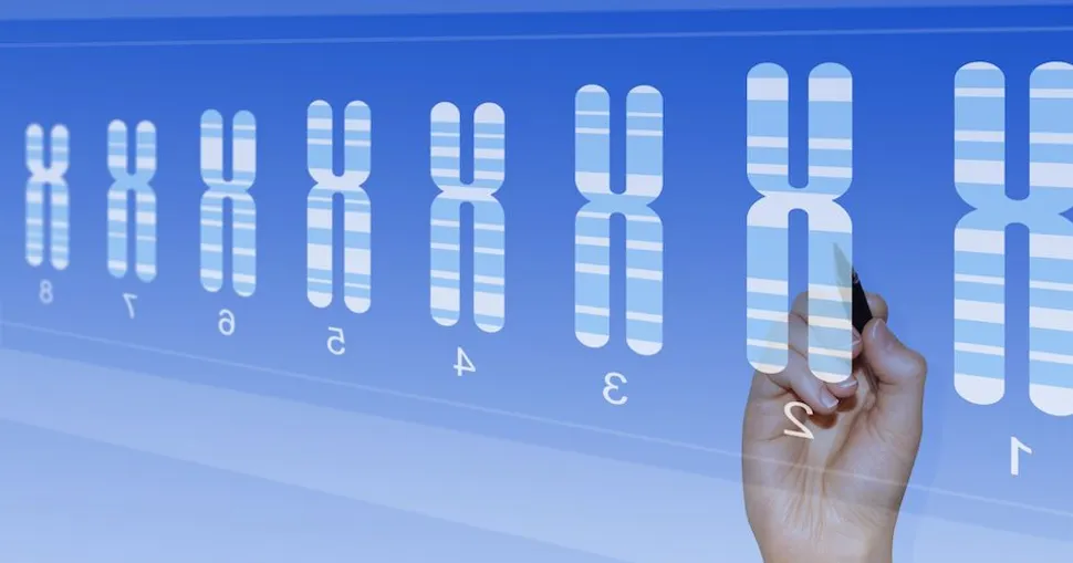
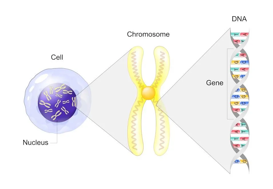
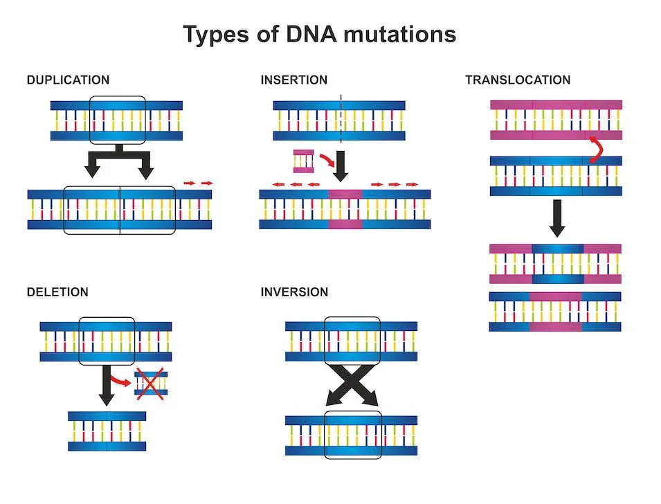
A CpG-stimulated karyotype test checks a patient’s chromosome for deletions, additions, and re-arranged parts (translocations). Chromosomes contain genetic instructions which tell the cell how to work.
(Image source: Shutterstock)
Chromosome abnormalities are believed to be caused by sources such as a virus, excess oxidative stress, errors during cell division, or are genetically inherited. The abnormal chromosome with altered cell instructions causes the cell to not work as it should.
Finding these chromosome abnormalities using a CpG-stimulated karyotype test can help predict how CLL cells will progress and what their reaction may be to different CLL treatments.
CpG refers to a cytosine and guanine nucleotide sequence found in DNA. The CpG oligonucleotides stimulate DNA methylation.
Methylation means - “A chemical reaction in the body in which a small molecule called a methyl group gets added to DNA, proteins, or other molecules. The addition of methyl groups can affect how some molecules act in the body." (cancer.gov).
Stimulating DNA methylation helps enhance the ability to detect subtle chromosomal abnormalities.
See here for chromosome abnormalities relevant to CLL.
(Image source: Shutterstock)
Steps of the Test
- Sample Collection:
- A small blood sample is taken from the patient by a trained healthcare professional or phlebotomist
- Cell Culturing:
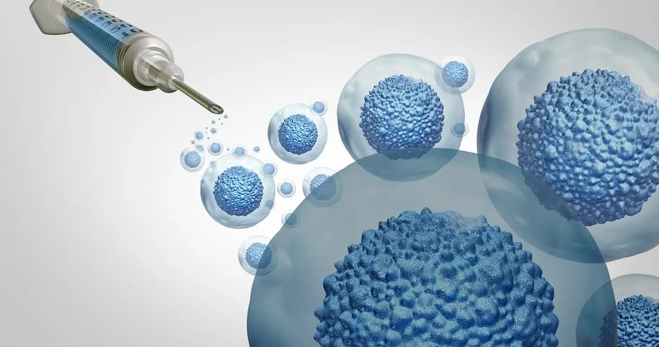
- In the laboratory, the collected blood sample is processed to extract and culture the patient's white blood cells, commonly lymphocytes. These cells are then treated with a solution that contains CpG oligonucleotides. The CpG oligonucleotides stimulate DNA methylation, helping show chromosomal abnormalities
- Stimulation and Chromosome Spreading:
- The CpG-stimulated cells are incubated to allow DNA methylation to occur. After the stimulation period, the cells are treated with a hypotonic solution, which causes the cells to swell and burst. This step, known as hypotonic shock, helps the chromosomes spread, making them more visible to analyze
- Chromosome Staining and Analysis:
- Once the chromosomes are adequately spread, they are stained using dyes, such as Giemsa or Wright stains. These stains provide a contrast that allows for better visualization of the chromosomes. Highly trained laboratory professionals then examine the stained chromosomes using a microscope and analyze their number, size, shape, and any structural abnormalities
Ask your CLL specialist about any questions you may have related to medical tests. Need help finding a CLL specialist? Check out HealthTree’s CLL specialist directory here.
What is a CpG-Stimulated Karyotype Test?
A CpG-stimulated karyotype test checks a patient’s chromosome for deletions, additions, and re-arranged parts (translocations). Chromosomes contain genetic instructions which tell the cell how to work.
(Image source: Shutterstock)
Chromosome abnormalities are believed to be caused by sources such as a virus, excess oxidative stress, errors during cell division, or are genetically inherited. The abnormal chromosome with altered cell instructions causes the cell to not work as it should.
Finding these chromosome abnormalities using a CpG-stimulated karyotype test can help predict how CLL cells will progress and what their reaction may be to different CLL treatments.
CpG refers to a cytosine and guanine nucleotide sequence found in DNA. The CpG oligonucleotides stimulate DNA methylation.
Methylation means - “A chemical reaction in the body in which a small molecule called a methyl group gets added to DNA, proteins, or other molecules. The addition of methyl groups can affect how some molecules act in the body." (cancer.gov).
Stimulating DNA methylation helps enhance the ability to detect subtle chromosomal abnormalities.
See here for chromosome abnormalities relevant to CLL.
(Image source: Shutterstock)
Steps of the Test
- Sample Collection:
- A small blood sample is taken from the patient by a trained healthcare professional or phlebotomist
- Cell Culturing:
- In the laboratory, the collected blood sample is processed to extract and culture the patient's white blood cells, commonly lymphocytes. These cells are then treated with a solution that contains CpG oligonucleotides. The CpG oligonucleotides stimulate DNA methylation, helping show chromosomal abnormalities
- Stimulation and Chromosome Spreading:
- The CpG-stimulated cells are incubated to allow DNA methylation to occur. After the stimulation period, the cells are treated with a hypotonic solution, which causes the cells to swell and burst. This step, known as hypotonic shock, helps the chromosomes spread, making them more visible to analyze
- Chromosome Staining and Analysis:
- Once the chromosomes are adequately spread, they are stained using dyes, such as Giemsa or Wright stains. These stains provide a contrast that allows for better visualization of the chromosomes. Highly trained laboratory professionals then examine the stained chromosomes using a microscope and analyze their number, size, shape, and any structural abnormalities
Ask your CLL specialist about any questions you may have related to medical tests. Need help finding a CLL specialist? Check out HealthTree’s CLL specialist directory here.

about the author
Megan Heaps
Megan joined HealthTree in 2022. She enjoys helping patients and their care partners understand the various aspects of the cancer. This understanding enables them to better advocate for themselves and improve their treatment outcomes.
More on Navigating Your Health


Trending Articles


Get the Latest Chronic Lymphocytic Leukemia Updates, Delivered to You.
By subscribing to the HealthTree newsletter, you'll receive the latest research, treatment updates, and expert insights to help you navigate your health.
Together we care.
Together we cure.