How is Large B Cell Lymphoma Diagnosed?
Large B-cell lymphoma (LBCL) is diagnosed through a combination of a physical examination, blood tests, lymph node biopsies, and imaging tests. The results obtained from these tests help determine if you have LBCL, what subtype of LBCL you have, and your treatment plan.
What are the Blood Tests that Detect LBCL?
-
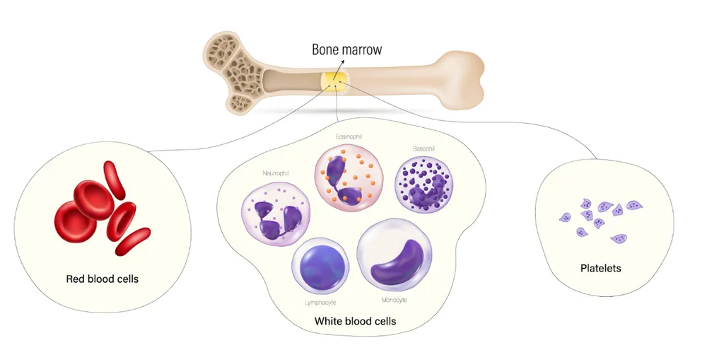
Complete blood count (CBC): A CBC measures the number of different types of blood cells, including red blood cells, white blood cells, and platelets. Abnormalities in the CBC can provide important diagnostic clues. In LBCL, the following CBC findings may be observed:
-
Anemia: LBCL can lead to anemia, characterized by a low red blood cell count and decreased hemoglobin levels, resulting in symptoms such as fatigue and weakness.
-
Leukocytosis: Elevated white blood cell counts, particularly an increased number of lymphocytes and monocytes, may be present in LBCL.
-
Thrombocytopenia: Low platelet counts can occur in LBCL, leading to a higher risk of bleeding and easy bruising.
-
Lactate dehydrogenase (LDH): LDH is an enzyme that is often elevated in the blood when there is cell damage or increased cell turnover, as seen in LBCL. Elevated LDH levels may be associated with a more aggressive disease.
-
Beta-2 microglobulin: Beta-2 microglobulin is a protein found on the surface of some immune cells. Elevated levels of beta-2 microglobulin can be associated with more advanced or aggressive lymphomas, including LBCL
Why are Lymph Nodes Analyzed?
Lymph nodes are one of the first signs of diffuse large B-cell lymphoma, therefore, it's important to analyze abnormal enlargement of lymph nodes.
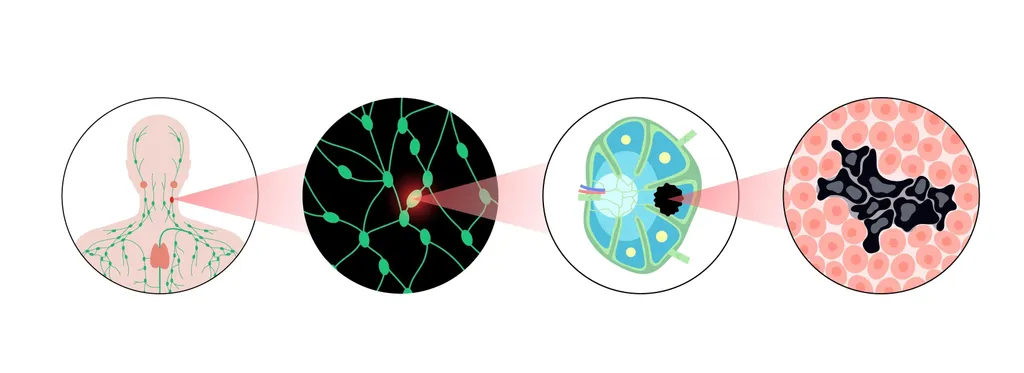
- Lymph node biopsy: The diagnosis of LBCL is confirmed by removing part or all of an enlarged lymph node with a biopsy. This procedure may be performed with local anesthesia if the involved tissue is relatively close to the skin's surface. If the node is deeper, general anesthesia is required. The cells from the tissue are then examined in detail using a microscope and other techniques.
- Immunohistochemistry: This test involves a staining process of the biopsy sample that adds a chemical marker to immune cells. The cells are then studied under a microscope.
- Molecular testing: The biopsy sample will undergo tests to look for specific DNA mutations and protein levels. Examples of proteins found in LBCL cells include BCL2, BCL6, CD3, CD5, CD10, CD20, CD45, IRF4/MUM1, Ki-67, and MYC.
Does LBCL Diagnosis Require Bone Marrow Tests?
Yes, the bone marrow is affected in 11–25% of DLBCL patients, but in advanced stages it can affect up to 60% of patients, so it's important to find out if, besides a lymph node, the LBCL cells affect the bone marrow.
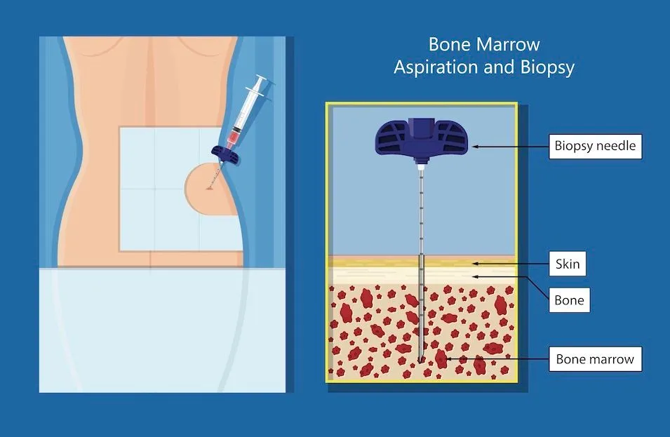
- Bone marrow aspiration and biopsy: These tests involve taking a small amount of bone marrow, usually from the hip bone, and examining it under a microscope to look for lymphoma cells. A bone marrow biopsy is unnecessary if a PET/CT scan shows the bone is affected.
- Flow cytometry: Flow cytometry is used to analyze the cell surface markers and characteristics of the cells present in the bone marrow. It can help identify and quantify the lymphoma cells and assess their immunophenotype.
- Cytogenetic analysis: Cytogenetic analysis involves the examination of the chromosomes within the bone marrow cells. It can detect chromosomal abnormalities or genetic mutations that may be associated with LBCL
- Molecular testing: Molecular testing may be performed to identify specific genetic mutations or rearrangements associated with LBCL. This testing can provide additional diagnostic and prognostic information.
- Fluorescence in situ hybridization (FISH): This test finds changes in chromosomes called translocations. Since this test doesn't need growing cells, it can be done on a bone marrow sample or a blood sample. In LBCL, FISH is used to detect MYC, BCL2, and BCL6 gene rearrangements. BCL6 rearrangement is the most common chromosomal abnormality found in DLBCL.
What are the Imaging Tests for LBCL?
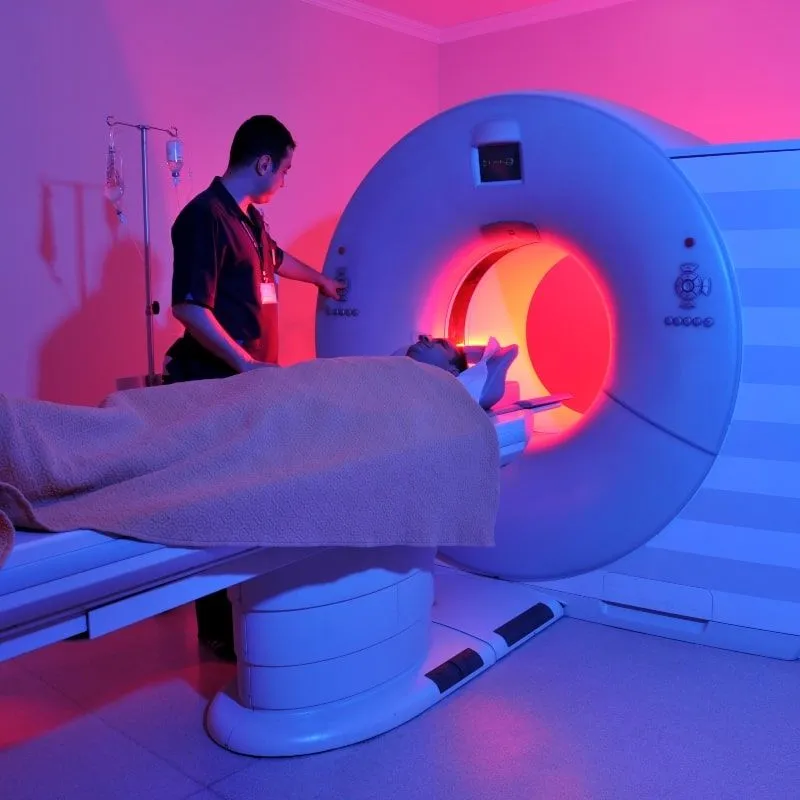
- Computed Tomography (CT) scan: This test uses X-rays to make detailed pictures of sections of the body, such as the chest, abdomen, and pelvis, where lymphoma often occurs.
- Positron Emission Tomography (PET) scan: This test uses a small amount of radioactive glucose (sugar) to find cancer cells in the body. The areas where the glucose is taken up appear 'hot' on the scan and can indicate the presence of lymphoma.
- Magnetic Resonance Imaging (MRI): This test uses magnetic fields, not x-rays, to produce detailed images of the body. It can be used to measure the size of the lymphoma.
It's important to note that the diagnosis and classification of LBCL are complex and require a comprehensive evaluation by a team of specialists. A complete diagnosis requires a physical examination, analysis of a bone marrow sample, and genetic testing.
The specific markers and genetic changes found in the bone marrow sample can also help determine a person's prognosis and guide treatment decisions. If you don't currently have an LBCL specialist on your team, it is important that you consult with one. Use HealthTree's LBCL Specialist Directory to locate a specialist near you.
Stay Informed with HealthTree News
Educated and empowered patients have better outcomes. That's why we want to help you learn more about your disease with the latest lymphoma news, including patient stories, treatment breakthroughs, and expert insights so you can make confident, informed decisions.
Large B-cell lymphoma (LBCL) is diagnosed through a combination of a physical examination, blood tests, lymph node biopsies, and imaging tests. The results obtained from these tests help determine if you have LBCL, what subtype of LBCL you have, and your treatment plan.
What are the Blood Tests that Detect LBCL?
-
Complete blood count (CBC): A CBC measures the number of different types of blood cells, including red blood cells, white blood cells, and platelets. Abnormalities in the CBC can provide important diagnostic clues. In LBCL, the following CBC findings may be observed:
-
Anemia: LBCL can lead to anemia, characterized by a low red blood cell count and decreased hemoglobin levels, resulting in symptoms such as fatigue and weakness.
-
Leukocytosis: Elevated white blood cell counts, particularly an increased number of lymphocytes and monocytes, may be present in LBCL.
-
Thrombocytopenia: Low platelet counts can occur in LBCL, leading to a higher risk of bleeding and easy bruising.
-
-
Lactate dehydrogenase (LDH): LDH is an enzyme that is often elevated in the blood when there is cell damage or increased cell turnover, as seen in LBCL. Elevated LDH levels may be associated with a more aggressive disease.
-
Beta-2 microglobulin: Beta-2 microglobulin is a protein found on the surface of some immune cells. Elevated levels of beta-2 microglobulin can be associated with more advanced or aggressive lymphomas, including LBCL
Why are Lymph Nodes Analyzed?
Lymph nodes are one of the first signs of diffuse large B-cell lymphoma, therefore, it's important to analyze abnormal enlargement of lymph nodes.
- Lymph node biopsy: The diagnosis of LBCL is confirmed by removing part or all of an enlarged lymph node with a biopsy. This procedure may be performed with local anesthesia if the involved tissue is relatively close to the skin's surface. If the node is deeper, general anesthesia is required. The cells from the tissue are then examined in detail using a microscope and other techniques.
- Immunohistochemistry: This test involves a staining process of the biopsy sample that adds a chemical marker to immune cells. The cells are then studied under a microscope.
- Molecular testing: The biopsy sample will undergo tests to look for specific DNA mutations and protein levels. Examples of proteins found in LBCL cells include BCL2, BCL6, CD3, CD5, CD10, CD20, CD45, IRF4/MUM1, Ki-67, and MYC.
Does LBCL Diagnosis Require Bone Marrow Tests?
Yes, the bone marrow is affected in 11–25% of DLBCL patients, but in advanced stages it can affect up to 60% of patients, so it's important to find out if, besides a lymph node, the LBCL cells affect the bone marrow.
- Bone marrow aspiration and biopsy: These tests involve taking a small amount of bone marrow, usually from the hip bone, and examining it under a microscope to look for lymphoma cells. A bone marrow biopsy is unnecessary if a PET/CT scan shows the bone is affected.
- Flow cytometry: Flow cytometry is used to analyze the cell surface markers and characteristics of the cells present in the bone marrow. It can help identify and quantify the lymphoma cells and assess their immunophenotype.
- Cytogenetic analysis: Cytogenetic analysis involves the examination of the chromosomes within the bone marrow cells. It can detect chromosomal abnormalities or genetic mutations that may be associated with LBCL
- Molecular testing: Molecular testing may be performed to identify specific genetic mutations or rearrangements associated with LBCL. This testing can provide additional diagnostic and prognostic information.
- Fluorescence in situ hybridization (FISH): This test finds changes in chromosomes called translocations. Since this test doesn't need growing cells, it can be done on a bone marrow sample or a blood sample. In LBCL, FISH is used to detect MYC, BCL2, and BCL6 gene rearrangements. BCL6 rearrangement is the most common chromosomal abnormality found in DLBCL.
What are the Imaging Tests for LBCL?
- Computed Tomography (CT) scan: This test uses X-rays to make detailed pictures of sections of the body, such as the chest, abdomen, and pelvis, where lymphoma often occurs.
- Positron Emission Tomography (PET) scan: This test uses a small amount of radioactive glucose (sugar) to find cancer cells in the body. The areas where the glucose is taken up appear 'hot' on the scan and can indicate the presence of lymphoma.
- Magnetic Resonance Imaging (MRI): This test uses magnetic fields, not x-rays, to produce detailed images of the body. It can be used to measure the size of the lymphoma.
It's important to note that the diagnosis and classification of LBCL are complex and require a comprehensive evaluation by a team of specialists. A complete diagnosis requires a physical examination, analysis of a bone marrow sample, and genetic testing.
The specific markers and genetic changes found in the bone marrow sample can also help determine a person's prognosis and guide treatment decisions. If you don't currently have an LBCL specialist on your team, it is important that you consult with one. Use HealthTree's LBCL Specialist Directory to locate a specialist near you.
Stay Informed with HealthTree News
Educated and empowered patients have better outcomes. That's why we want to help you learn more about your disease with the latest lymphoma news, including patient stories, treatment breakthroughs, and expert insights so you can make confident, informed decisions.
Trending Articles


Get the Latest Large B Cell Lymphoma Updates, Delivered to You.
By subscribing to the HealthTree newsletter, you'll receive the latest research, treatment updates, and expert insights to help you navigate your health.
Together we care.
Together we cure.