Lymphoma PET/CT Scan: How It Works and What Patients Can Expect
Learn what a PET/CT scan is, how it detects lymphoma, what to expect during and after the scan, and how doctors interpret the results.
What is a lymphoma PET/CT scan?
A PET/CT scan for lymphoma combines two imaging tests in one procedure: a positron emission tomography (PET) scan and a computed tomography (CT) scan.
PET scan vs CT scan for lymphoma
A PET scan shows how active cells are in the body. It highlights areas where cells use a lot of energy. These areas may appear as bright spots on the scan and can suggest lymphoma activity.
Before the scan, a small amount of a radioactive sugar called fluorodeoxyglucose (FDG) is injected into a vein. FDG works like glucose, the sugar your body uses for energy. Because lymphoma cells often use more sugar than normal cells, they absorb more FDG and appear brighter on the PET scan.
A CT scan, on the other hand, creates detailed images of organs and tissues. Patients may wonder if you can see lymphoma on a CT scan. In some cases, yes. A CT scan can show lymph nodes that look larger or different from normal. However, a CT scan does not show how active the cells are. Lymphoma can sometimes be present even when lymph nodes appear normal in size.
When doctors combine both scans in a PET/CT, they can see where lymphoma may be located and how active the cells are. This can help your care team better understand the lymphoma, plan the best treatment for you, or monitor how well a treatment is working.
How to prepare for a PET/CT scan
Preparing for a PET/CT scan helps ensure accurate results. Your care team will tell you exactly how they want you to prepare. You may need to:
- Avoid eating for 4 to 6 hours before the scan.
- Drink water but avoid sugary drinks.
- Avoid strenuous exercise the day before the scan.
- Inform your care team about medications or health conditions.
Blood sugar levels are often checked before the scan because high glucose levels can affect how the tracer works.
Wearing comfortable clothing and removing metal items or jewelry may also be recommended.
What to expect during and after the scan
A full PET/CT appointment usually takes about 1.5 to 2 hours.
First, a technologist injects the FDG tracer into a vein. After the injection, you will wait about 60 minutes while the tracer moves through the body.
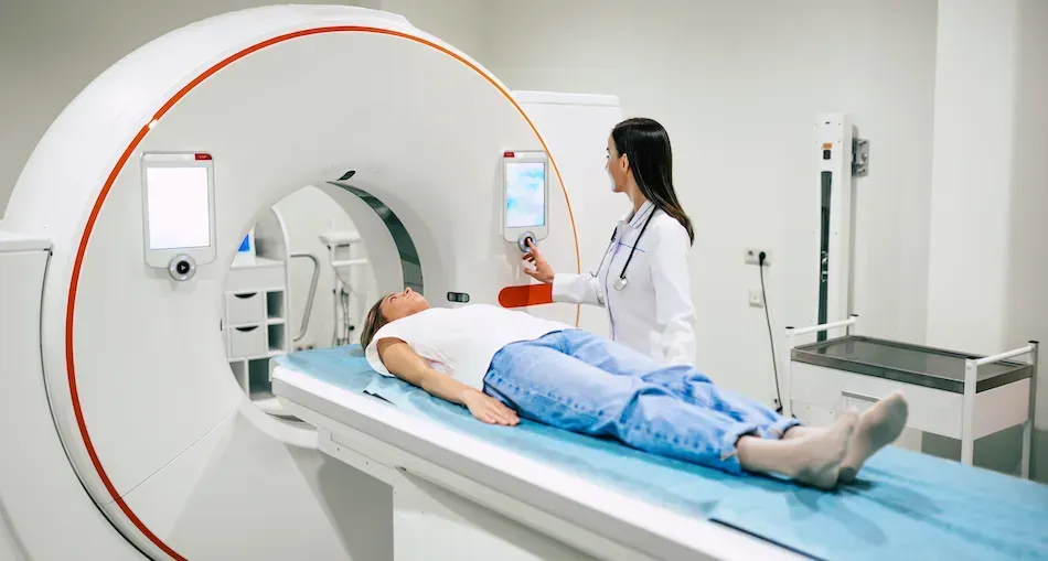
Next, you will lie on a table that slowly slides into the scanning machine. It is important to stay still while the images are taken. The scanning part usually takes about 30 minutes and is painless.
After the scan, most people can return to normal activities right away. The tracer gradually leaves the body through urine or stool over several hours or days. Drinking plenty of water can help the body clear the tracer faster.
How doctors interpret PET/CT results
A radiologist reviews the images and sends a report to your doctor. Results are often available within a few days.
Doctors frequently use a system called the Deauville five-point scale, which compares the activity seen on the scan with normal areas such as the liver.
- Scores 1 to 3 often mean there is little or no active lymphoma.
- Scores 4 to 5 may suggest remaining lymphoma or progression.
Your care team will review the results with you and explain what they mean for your care plan.
Benefits and possible risks of a PET/CT scan
A PET/CT scan provides several benefits for lymphoma care:
- More accurate staging of lymphoma
- Earlier detection of active lymphoma
- Ability to evaluate how well treatment is working
- Better information to guide treatment decisions
The possible risks are generally low but may include:
- Exposure to a small amount of radiation
- Rare reactions to the tracer or contrast material
- False positives caused by inflammation or infection
Doctors recommend a PET/CT scan when the benefits outweigh these risks.
Does a PET/CT scan work the same for all lymphoma subtypes?
A PET/CT scan is especially useful for aggressive lymphomas such as Hodgkin lymphoma and large B-cell lymphoma (LBCL) because these cancers often show strong tracer uptake.
Some slower-growing lymphomas may show less uptake on the scan. These may include:
In these cases, doctors may use a PET/CT scan alongside other imaging tests to monitor lymphoma.
Key takeaways for people living with lymphoma
A PET/CT scan helps doctors detect lymphoma, determine its stage, and evaluate how well treatment is working. By combining metabolic and structural imaging, this test provides a clearer picture of lymphoma activity throughout the body. Understanding what the scan shows and how it works can help you feel more prepared about the imaging tests part of your care.
Get the latest lymphoma updates delivered to you! The HealthTree newsletter shares core education, research advances, and more directly to your inbox.
Sources:
- Nuclear Medicine PET/CT Lymphomas Assessment, Protocols, and Interpretation
- Mapping the research landscape of PET/CT in lymphoma: insights from a bibliometric analysis
- PET-CT Imaging of Lymphoma
- Radiology PET/CT
- Effect of PET/CT on the Management and Outcomes of Participants with Hodgkin and Aggressive Non-Hodgkin Lymphoma: A Multicenter Registry
Learn what a PET/CT scan is, how it detects lymphoma, what to expect during and after the scan, and how doctors interpret the results.
What is a lymphoma PET/CT scan?
A PET/CT scan for lymphoma combines two imaging tests in one procedure: a positron emission tomography (PET) scan and a computed tomography (CT) scan.
PET scan vs CT scan for lymphoma
A PET scan shows how active cells are in the body. It highlights areas where cells use a lot of energy. These areas may appear as bright spots on the scan and can suggest lymphoma activity.
Before the scan, a small amount of a radioactive sugar called fluorodeoxyglucose (FDG) is injected into a vein. FDG works like glucose, the sugar your body uses for energy. Because lymphoma cells often use more sugar than normal cells, they absorb more FDG and appear brighter on the PET scan.
A CT scan, on the other hand, creates detailed images of organs and tissues. Patients may wonder if you can see lymphoma on a CT scan. In some cases, yes. A CT scan can show lymph nodes that look larger or different from normal. However, a CT scan does not show how active the cells are. Lymphoma can sometimes be present even when lymph nodes appear normal in size.
When doctors combine both scans in a PET/CT, they can see where lymphoma may be located and how active the cells are. This can help your care team better understand the lymphoma, plan the best treatment for you, or monitor how well a treatment is working.
How to prepare for a PET/CT scan
Preparing for a PET/CT scan helps ensure accurate results. Your care team will tell you exactly how they want you to prepare. You may need to:
- Avoid eating for 4 to 6 hours before the scan.
- Drink water but avoid sugary drinks.
- Avoid strenuous exercise the day before the scan.
- Inform your care team about medications or health conditions.
Blood sugar levels are often checked before the scan because high glucose levels can affect how the tracer works.
Wearing comfortable clothing and removing metal items or jewelry may also be recommended.
What to expect during and after the scan
A full PET/CT appointment usually takes about 1.5 to 2 hours.
First, a technologist injects the FDG tracer into a vein. After the injection, you will wait about 60 minutes while the tracer moves through the body.
Next, you will lie on a table that slowly slides into the scanning machine. It is important to stay still while the images are taken. The scanning part usually takes about 30 minutes and is painless.
After the scan, most people can return to normal activities right away. The tracer gradually leaves the body through urine or stool over several hours or days. Drinking plenty of water can help the body clear the tracer faster.
How doctors interpret PET/CT results
A radiologist reviews the images and sends a report to your doctor. Results are often available within a few days.
Doctors frequently use a system called the Deauville five-point scale, which compares the activity seen on the scan with normal areas such as the liver.
- Scores 1 to 3 often mean there is little or no active lymphoma.
- Scores 4 to 5 may suggest remaining lymphoma or progression.
Your care team will review the results with you and explain what they mean for your care plan.
Benefits and possible risks of a PET/CT scan
A PET/CT scan provides several benefits for lymphoma care:
- More accurate staging of lymphoma
- Earlier detection of active lymphoma
- Ability to evaluate how well treatment is working
- Better information to guide treatment decisions
The possible risks are generally low but may include:
- Exposure to a small amount of radiation
- Rare reactions to the tracer or contrast material
- False positives caused by inflammation or infection
Doctors recommend a PET/CT scan when the benefits outweigh these risks.
Does a PET/CT scan work the same for all lymphoma subtypes?
A PET/CT scan is especially useful for aggressive lymphomas such as Hodgkin lymphoma and large B-cell lymphoma (LBCL) because these cancers often show strong tracer uptake.
Some slower-growing lymphomas may show less uptake on the scan. These may include:
In these cases, doctors may use a PET/CT scan alongside other imaging tests to monitor lymphoma.
Key takeaways for people living with lymphoma
A PET/CT scan helps doctors detect lymphoma, determine its stage, and evaluate how well treatment is working. By combining metabolic and structural imaging, this test provides a clearer picture of lymphoma activity throughout the body. Understanding what the scan shows and how it works can help you feel more prepared about the imaging tests part of your care.
Get the latest lymphoma updates delivered to you! The HealthTree newsletter shares core education, research advances, and more directly to your inbox.
Sources:
- Nuclear Medicine PET/CT Lymphomas Assessment, Protocols, and Interpretation
- Mapping the research landscape of PET/CT in lymphoma: insights from a bibliometric analysis
- PET-CT Imaging of Lymphoma
- Radiology PET/CT
- Effect of PET/CT on the Management and Outcomes of Participants with Hodgkin and Aggressive Non-Hodgkin Lymphoma: A Multicenter Registry

about the author
Megan Heaps
Megan joined HealthTree in 2022. She enjoys helping patients and their care partners understand the various aspects of the cancer. This understanding enables them to better advocate for themselves and improve their treatment outcomes.
More on Core Education


Trending Articles


Get the Latest Large B Cell Lymphoma Updates, Delivered to You.
By subscribing to the HealthTree newsletter, you'll receive the latest research, treatment updates, and expert insights to help you navigate your health.
Together we care.
Together we cure.