Understanding Sarcoma

How is Sarcoma Diagnosed?
Last updated and reviewed on May 27, 2026.
Getting the right diagnosis is one of the most important steps when sarcoma is suspected. Because sarcoma is rare and can look like other conditions on imaging, it is very important to be evaluated by a team with experience in diagnosing and treating sarcoma. The diagnosis process usually involves a combination of imaging scans, a tissue sample (called a biopsy), and lab tests.
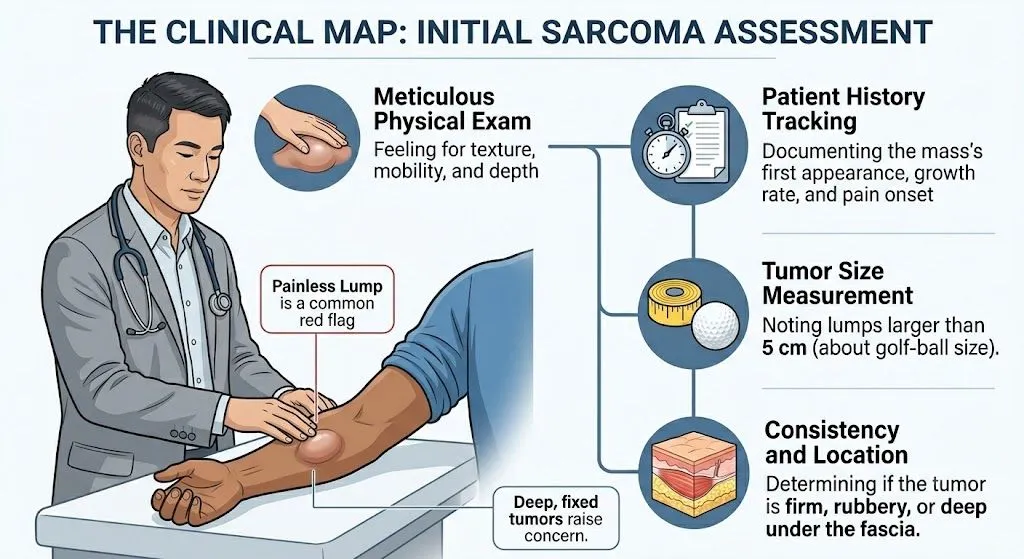
Step 1: Physical Exam and Medical History
Your doctor will start by asking about your symptoms, how long you have had them, whether they are getting worse, and whether you have any known risk factors. The doctor will also do a physical exam, feeling for any lumps or areas of tenderness.
Step 2: Imaging Tests
Imaging tests let doctors see inside the body without surgery. They are used to find the tumor, see how big it is, check whether it has grown into nearby structures, and look for signs that it has spread to other parts of the body (called metastasis).
- MRI (Magnetic Resonance Imaging) MRI is the most useful imaging test for soft tissue sarcomas. It uses magnetic fields and radio waves, not X-rays, to create detailed pictures of soft tissue. An MRI shows the size of the tumor, its exact location, and whether it is pressing on important structures like blood vessels or nerves. This information is crucial for planning surgery.
- CT Scan (Computed Tomography) A CT scan takes many X-ray images from different angles and puts them together into a detailed picture. CT scans are commonly used to look at tumors in the abdomen or chest, and to check whether the sarcoma has spread to the lungs (the most common site of spread for many sarcomas).
- X-Ray Plain X-rays are often the first test done when bone cancer is suspected. They can show abnormal areas in bones, destruction of bone tissue, or unusual new bone formation, all of which can be signs of a bone sarcoma.
- PET Scan (Positron Emission Tomography) A PET scan shows how active cells are in the body. Because cancer cells use more energy (sugar/glucose) than normal cells, they show up brightly on a PET scan. PET scans are sometimes used to see if the sarcoma has spread to lymph nodes or distant organs.
- Bone Scan A bone scan uses a small amount of radioactive material to find areas of abnormal bone activity. It can help determine whether bone sarcoma has spread to other bones.
Step 3: Biopsy
A biopsy is the only way to know for certain whether a tumor is cancer. In a biopsy, a small piece of tissue is removed from the tumor and looked at under a microscope by a pathologist (a doctor who specializes in studying cells and tissues).
Why biopsy planning matters so much. For sarcoma, the biopsy must be done very carefully and should be planned by the surgeon who will perform the main surgery. A poorly placed biopsy incision (cut) can contaminate healthy tissue with cancer cells, making future surgery more complicated or even impossible to perform safely. This is one of the most important reasons to seek care at a sarcoma specialty center before having a biopsy.
There are several types of biopsy:
- Core needle biopsy: A hollow needle is inserted through the skin into the tumor to remove a small core of tissue. This is the most common type used for sarcoma and is usually done with imaging guidance (like ultrasound or CT) to make sure the needle goes to the right place.
- Incisional biopsy: A surgeon makes a small cut and removes a piece of the tumor. This is sometimes needed when a needle biopsy does not provide enough tissue.
- Excisional biopsy: The entire lump is removed for examination. This is generally not recommended for suspected sarcoma unless the tumor is very small, because it can make future surgery more difficult.
Questions to Ask Before Your Biopsy
If your doctor suggests a biopsy for a suspicious lump, you are the final checkpoint for your safety. Ask these five questions before allowing a procedure to proceed:
-
Is your team a dedicated sarcoma multidisciplinary team (MDT), or are you a general oncologist/surgeon? (The difference can dramatically impact your long-term outcome.)
-
How will you ensure that the biopsy needle track doesn't contaminate healthy muscle tissue that I will need for reconstruction after surgery? (A general surgeon might place a needle track carelessly; a sarcoma surgeon will place it precisely along the intended future surgical incision.)
-
Are you planning to use an image-guided core needle biopsy (the standard for sarcoma) or are you planning to do an incisional/excisional biopsy? (Removing the whole lump—an excisional biopsy—can make curative surgery vastly more complex later on.)
Will the surgeon who will be performing my final tumor removal surgery be the one planning the exact trajectory of this biopsy needle?
Will this tissue sample be reviewed by a specialist pathologist who primarily diagnoses sarcoma? (Mesenchymal tumors are notoriously difficult to subtype, and a general pathologist may misclassify the cancer.)
Step 4: Lab Tests on the Tumor Tissue
Once the biopsy tissue is collected, the pathologist performs several tests:
- Histology: The tissue is examined under a microscope to see what kind of cells make up the tumor. This tells doctors the type of sarcoma and its grade (how abnormal the cells look).
- Immunohistochemistry (IHC) Special stains are applied to the tissue to look for certain proteins on the cancer cells. This helps identify the specific subtype of sarcoma.
- Molecular and Genetic Testing Many sarcomas have specific gene changes (mutations or translocations) that can be detected with molecular tests. For example, Ewing sarcoma has a characteristic EWSR1 gene fusion, and GISTs often have KIT or PDGFRA mutations. These test results can confirm the diagnosis and sometimes guide treatment choices (for example, targeted therapy for GISTs).
Step 5: Blood Tests
Blood tests cannot diagnose sarcoma, but they are used to check your overall health and organ function before starting treatment. Your doctor may check your blood counts, kidney function, liver function, and other measures to make sure you are healthy enough for surgery or chemotherapy.
The Importance of a Specialist Team
Because sarcoma is so rare and complex, getting a diagnosis from an experienced sarcoma team is critical. A multidisciplinary team (MDT), meaning a group of specialists including a surgical oncologist, medical oncologist, radiation oncologist, radiologist, and pathologist, all with sarcoma experience, should be involved in reviewing your case. Studies show that patients treated at sarcoma specialty centers have better outcomes than those treated at centers without sarcoma expertise.
|
What’s Next: The next page in this guide is How is Sarcoma Staged and Classified?. If you would like to read another page in this guide, return to the Sarcoma 101 Guides page or choose another topic. |
Sources:
-
American Cancer Society. Soft Tissue Sarcoma: Tests for Soft Tissue Sarcomas. https://www.cancer.org/cancer/types/soft-tissue-sarcoma/detection-diagnosis-staging/how-diagnosed.html
National Cancer Institute. Soft Tissue Sarcoma Treatment (PDQ) – Patient Version. https://www.cancer.gov/types/soft-tissue-sarcoma/patient/adult-soft-tissue-treatment-pdq
Kaste SC. Issues specific to implementing PET-CT for pediatric oncology: what we have learned from childhood sarcoma studies. Pediatric Radiology. 2004. https://pubmed.ncbi.nlm.nih.gov/14745525/
Grimer R, et al. Guidelines for the management of soft tissue sarcomas. Sarcoma. 2010;2010:506182. https://pubmed.ncbi.nlm.nih.gov/20634933/
Von Mehren M, et al. NCCN Clinical Practice Guidelines in Oncology: Soft Tissue Sarcoma. Journal of the National Comprehensive Cancer Network. 2022. https://pubmed.ncbi.nlm.nih.gov/35830886/
How is Sarcoma Diagnosed?
Last updated and reviewed on May 27, 2026.
Getting the right diagnosis is one of the most important steps when sarcoma is suspected. Because sarcoma is rare and can look like other conditions on imaging, it is very important to be evaluated by a team with experience in diagnosing and treating sarcoma. The diagnosis process usually involves a combination of imaging scans, a tissue sample (called a biopsy), and lab tests.
Step 1: Physical Exam and Medical History
Your doctor will start by asking about your symptoms, how long you have had them, whether they are getting worse, and whether you have any known risk factors. The doctor will also do a physical exam, feeling for any lumps or areas of tenderness.
Step 2: Imaging Tests
Imaging tests let doctors see inside the body without surgery. They are used to find the tumor, see how big it is, check whether it has grown into nearby structures, and look for signs that it has spread to other parts of the body (called metastasis).
- MRI (Magnetic Resonance Imaging) MRI is the most useful imaging test for soft tissue sarcomas. It uses magnetic fields and radio waves, not X-rays, to create detailed pictures of soft tissue. An MRI shows the size of the tumor, its exact location, and whether it is pressing on important structures like blood vessels or nerves. This information is crucial for planning surgery.
- CT Scan (Computed Tomography) A CT scan takes many X-ray images from different angles and puts them together into a detailed picture. CT scans are commonly used to look at tumors in the abdomen or chest, and to check whether the sarcoma has spread to the lungs (the most common site of spread for many sarcomas).
- X-Ray Plain X-rays are often the first test done when bone cancer is suspected. They can show abnormal areas in bones, destruction of bone tissue, or unusual new bone formation, all of which can be signs of a bone sarcoma.
- PET Scan (Positron Emission Tomography) A PET scan shows how active cells are in the body. Because cancer cells use more energy (sugar/glucose) than normal cells, they show up brightly on a PET scan. PET scans are sometimes used to see if the sarcoma has spread to lymph nodes or distant organs.
- Bone Scan A bone scan uses a small amount of radioactive material to find areas of abnormal bone activity. It can help determine whether bone sarcoma has spread to other bones.
Step 3: Biopsy
A biopsy is the only way to know for certain whether a tumor is cancer. In a biopsy, a small piece of tissue is removed from the tumor and looked at under a microscope by a pathologist (a doctor who specializes in studying cells and tissues).
Why biopsy planning matters so much. For sarcoma, the biopsy must be done very carefully and should be planned by the surgeon who will perform the main surgery. A poorly placed biopsy incision (cut) can contaminate healthy tissue with cancer cells, making future surgery more complicated or even impossible to perform safely. This is one of the most important reasons to seek care at a sarcoma specialty center before having a biopsy.
There are several types of biopsy:
- Core needle biopsy: A hollow needle is inserted through the skin into the tumor to remove a small core of tissue. This is the most common type used for sarcoma and is usually done with imaging guidance (like ultrasound or CT) to make sure the needle goes to the right place.
- Incisional biopsy: A surgeon makes a small cut and removes a piece of the tumor. This is sometimes needed when a needle biopsy does not provide enough tissue.
- Excisional biopsy: The entire lump is removed for examination. This is generally not recommended for suspected sarcoma unless the tumor is very small, because it can make future surgery more difficult.
Questions to Ask Before Your Biopsy
If your doctor suggests a biopsy for a suspicious lump, you are the final checkpoint for your safety. Ask these five questions before allowing a procedure to proceed:
-
Is your team a dedicated sarcoma multidisciplinary team (MDT), or are you a general oncologist/surgeon? (The difference can dramatically impact your long-term outcome.)
-
How will you ensure that the biopsy needle track doesn't contaminate healthy muscle tissue that I will need for reconstruction after surgery? (A general surgeon might place a needle track carelessly; a sarcoma surgeon will place it precisely along the intended future surgical incision.)
-
Are you planning to use an image-guided core needle biopsy (the standard for sarcoma) or are you planning to do an incisional/excisional biopsy? (Removing the whole lump—an excisional biopsy—can make curative surgery vastly more complex later on.)
-
Will the surgeon who will be performing my final tumor removal surgery be the one planning the exact trajectory of this biopsy needle?
-
Will this tissue sample be reviewed by a specialist pathologist who primarily diagnoses sarcoma? (Mesenchymal tumors are notoriously difficult to subtype, and a general pathologist may misclassify the cancer.)
Step 4: Lab Tests on the Tumor Tissue
Once the biopsy tissue is collected, the pathologist performs several tests:
- Histology: The tissue is examined under a microscope to see what kind of cells make up the tumor. This tells doctors the type of sarcoma and its grade (how abnormal the cells look).
- Immunohistochemistry (IHC) Special stains are applied to the tissue to look for certain proteins on the cancer cells. This helps identify the specific subtype of sarcoma.
- Molecular and Genetic Testing Many sarcomas have specific gene changes (mutations or translocations) that can be detected with molecular tests. For example, Ewing sarcoma has a characteristic EWSR1 gene fusion, and GISTs often have KIT or PDGFRA mutations. These test results can confirm the diagnosis and sometimes guide treatment choices (for example, targeted therapy for GISTs).
Step 5: Blood Tests
Blood tests cannot diagnose sarcoma, but they are used to check your overall health and organ function before starting treatment. Your doctor may check your blood counts, kidney function, liver function, and other measures to make sure you are healthy enough for surgery or chemotherapy.
The Importance of a Specialist Team
Because sarcoma is so rare and complex, getting a diagnosis from an experienced sarcoma team is critical. A multidisciplinary team (MDT), meaning a group of specialists including a surgical oncologist, medical oncologist, radiation oncologist, radiologist, and pathologist, all with sarcoma experience, should be involved in reviewing your case. Studies show that patients treated at sarcoma specialty centers have better outcomes than those treated at centers without sarcoma expertise.
|
What’s Next: The next page in this guide is How is Sarcoma Staged and Classified?. If you would like to read another page in this guide, return to the Sarcoma 101 Guides page or choose another topic. |
Sources:
-
American Cancer Society. Soft Tissue Sarcoma: Tests for Soft Tissue Sarcomas. https://www.cancer.org/cancer/types/soft-tissue-sarcoma/detection-diagnosis-staging/how-diagnosed.html
-
National Cancer Institute. Soft Tissue Sarcoma Treatment (PDQ) – Patient Version. https://www.cancer.gov/types/soft-tissue-sarcoma/patient/adult-soft-tissue-treatment-pdq
-
Kaste SC. Issues specific to implementing PET-CT for pediatric oncology: what we have learned from childhood sarcoma studies. Pediatric Radiology. 2004. https://pubmed.ncbi.nlm.nih.gov/14745525/
-
Grimer R, et al. Guidelines for the management of soft tissue sarcomas. Sarcoma. 2010;2010:506182. https://pubmed.ncbi.nlm.nih.gov/20634933/
-
Von Mehren M, et al. NCCN Clinical Practice Guidelines in Oncology: Soft Tissue Sarcoma. Journal of the National Comprehensive Cancer Network. 2022. https://pubmed.ncbi.nlm.nih.gov/35830886/
Trending Articles


Get the Latest Solid Tumors Updates, Delivered to You.
By subscribing to the HealthTree newsletter, you'll receive the latest research, treatment updates, and expert insights to help you navigate your health.
Together we care.
Together we cure.